UVEA & OCULAR INFLAMMATION

The eye consists of the following basic layers :

Fibrous (outer) layer—the sclera and cornea

Vascular (middle) layer—the uvea, or uveal tract

Neuroectodermal inner layer—the retina and optic nerve.


The uveal tract has three parts: the iris and the ciliary body, which together form the anterior uvea, and the choroid, which is also known as the posterior uvea.

The following terms describe inflammation of the various parts of the uveal parts :

Different types of uveitis affect different parts of the eye.

  • TYPE - ANTERIOR UVEITIS

    Also called iritis or iridocyclitis, anterior uveitis is the most common form of uveitis. It is characterized by inflammation of the front of the eye, between the back of the cornea and in front of the lens. Anterior uveitis can be present in one or both eyes. Symptoms of this form of uveitis may include eye pain, red eyes, blurred vision and sensitivity to light.

  • TYPE - INTERMEDIATE UVEITIS

    Also called cyclitis or vitritis, intermediate uveitis is an intraocular inflammation primarily affecting the vitreous, which is the gel in the middle of the eye and behind the lens. Symptoms of intermediate uveitis may include blurred vision and spots in vision (commonly called floaters).

  • TYPE - POSTERIOR UVEITIS

    Also known as retinitis or choroiditis, posterior uveitis is the inflammation of the back of the eye. This primarily affects the retina, which includes the retinal vessels, or the choroid. Posterior uveitis is known to cause vision loss and may include symptoms of flashing lights or floaters.

  • TYPE - PANUVEITIS

    Panuveitis involves inflammation inside the eye that similarly affects the front, middle and back of the eye. It may be associated with any of the symptoms described in the other types of uveitis.

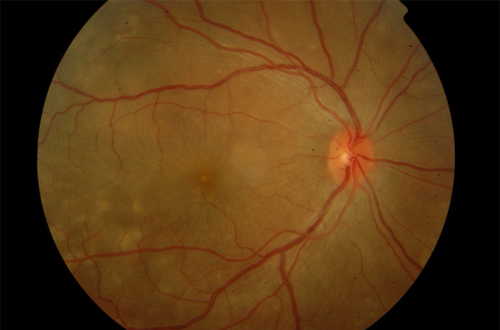
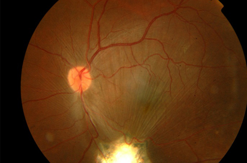
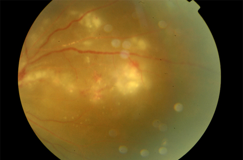
    Because of the continuity between the various parts of the uvea, aqueous humor, and vitreous, however, uveal inflammation often involves many ocular structures. The retina and choroid are adjacent, with no major barriers between, so they are frequently inflamed together.

    Clinical signs more specific for uveitis are as follows:

    • Aqueous flare
    • Inflammatory cells free in the anterior
    • chamber or adherent to the corneal
    • endothelium (keratic precipitates)
    • Hypopyon or hyphema
    • Episcleral vascular injection or
    • circumcorneal ciliary flush
    • Corneal edema
    • Miosis
    • Resistance to mydriatics
    • Lowered IOP
    • Anterior or posterior synechiae
    • Swollen or dull appearance of the iris
    • Increased pigmentation of the iris
    • Vitreous haze or opacity
    • Retinal edema, exudate, or detachment
    • Aqueous lipemia, which may be seen if
    • circulating lipid levels are high.

WHAT ARE THE INVESTIGATIONS FOR UVEITIS?

Ocular investigations include fundus fluorescein angiography, optical coherence tomography, B scan ultrasound and UBM.Systemic investigations include blood tests and radiological imaging.

IS STEROID THE ONLY TREATMENT FOR UVEITIS?

  • Infectious uveitis is treated with appropriate antibiotics or antivirals. Non infectious uveitis is treated with steroids. If prolonged treatment is required or if the patient cannot tolerate steroids, steroid sparing agents can be used. Patients will be regularly monitored for the adverse effects of the medicines and the treatment will be altered if any adverse effects develop.
  • Regular follow-up and strict allegiance to treatment is a must for all patients with uveitis.
  • Self medication can lead to complications. Hence avoid self medication and irregular treatment.


Get Appointment - If You Need Consultation

CONTACT DETAILS